DEXA Body Composition Scan: What to Expect, How it Works, & Why it Matters
Evaluating a patient’s health based solely on total body weight or Body Mass Index (BMI) often falls short of providing a complete metabolic profile. As healthcare providers, you understand that two patients with the same BMI can possess vastly different metabolic risk factors and physical capabilities. Consequently, there is a growing clinical requirement for objective, highly accurate tissue quantification.
Used as a clinical gold standard, a
DEXA body composition scan provides practitioners with a look into a patient's exact musculoskeletal and adipose tissue distribution. This comprehensive guide outlines the mechanisms behind Dual-Energy X-ray Absorptiometry (DEXA), what patients can expect during the procedure, how to interpret the diagnostic output, and why this data matters for proactive patient care.
What is a DEXA Body Composition Scan?
A DEXA body composition scan is a non-invasive, low-exposure radiographic imaging modality. While it has historically been used almost exclusively for bone densitometry, modern advancements have positioned the DEXA scan for body composition as a premier instrument for three-compartment tissue analysis. Unlike traditional assessment tools, it segments total body mass into three distinct categories: fat mass, lean soft tissue, and bone mineral content.
When advising patients, it is helpful to contrast this technology with other common methods. BMI remains a tool that fails to differentiate between high muscle mass and adiposity. Skinfold callipers show significant inter-operator variability and assess only subcutaneous fat. Similarly, bioelectrical impedance analysis (BIA) is heavily influenced by a patient’s acute hydration levels, food intake, and skin temperature, often yielding inconsistent longitudinal data.
A DEXA scan circumvents these limitations, offering clinicians stable, reproducible, and highly accurate baseline data.
How Does a DEXA Scan Work for Body Composition?
To understand how a body composition scan works, one must look at the underlying physics of differential attenuation. The equipment emits two distinct, low-energy X-ray beams through the patient's body tissues. Bone mineral, lean muscle tissue, and adipose fat tissue each attenuate (absorb) these X-ray beams differently.
When considering how a DEXA scan works for body composition, specialised detectors measure the exact amount of radiation passing through the body at each energy level. Software processes these values pixel-by-pixel, calculating the precise mass and percentage of each tissue compartment. The radiation exposure is remarkably low, often equivalent to less than one day of natural background radiation, making it a safe diagnostic tool for routine monitoring.
What to Expect: Preparing for a DEXA Body Composition Scan
When instructing patients on how to prepare for a DEXA body composition scan, simplicity and consistency are key. Patients should wear loose, comfortable clothing free of metal, as metal can disrupt the imaging. While patients do not need to alter their long-term diet, they should ideally fast from food and limit heavy fluid intake for 2 to 4 hours before their appointment to avoid transient gastrointestinal mass that could affect the trunk readings.
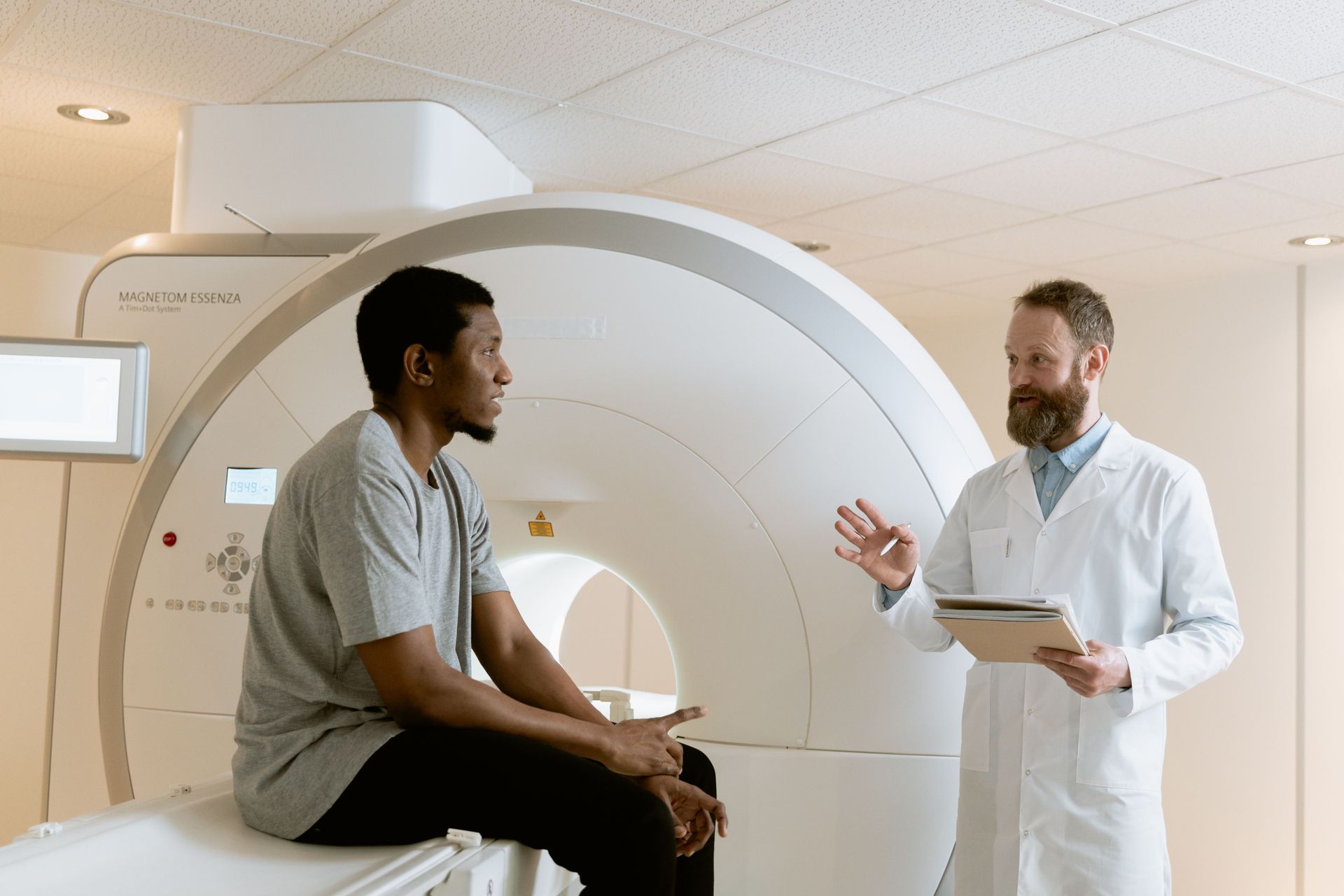
The actual scanning process is swift and entirely non-invasive. The patient lies supine and completely still on a padded, open scanner bed while the imaging arm slowly passes from head to toe. It takes approximately 6 to 10 minutes. Throughout the process, the patient remains fully clothed, experiences no physical discomfort, and can resume normal activities immediately.
Understanding Your Results: How to Read a DEXA Scan
Reviewing the comprehensive digital report allows clinicians to develop highly targeted, data-driven treatment plans. Knowing how to read DEXA scan results for body composition involves analysing several key parameters. The report breaks down tissue metrics globally and regionally across the android (abdominal), gynoid (hip/thigh), arms, legs, and trunk zones.
Key data points include:
- Total Body Fat Percentage: The overall ratio of adipose tissue to total body mass.
- Android/Gynoid (A/G) Ratio: A high A/G ratio indicates preferential fat deposition in the abdomen, a key marker of metabolic syndrome.
- Visceral Adipose Tissue (VAT): Quantifies the deep fat surrounding internal organs.
- Lean Mass Balance: Compares left-versus-right limb muscle mass to highlight functional asymmetries or atrophy.
Healthy reference ranges are adjusted for age and biological sex. Clinicians should view significant regional asymmetries or elevated VAT metrics as clinical "red flags" indicating elevated cardiovascular, metabolic, or biomechanical injury risks.
Bone Density and DEXA Scans
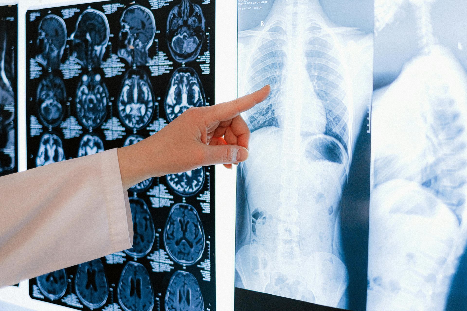
Beyond soft-tissue metrics, integrating a structural bone-density DEXA scan provides essential insights into skeletal integrity. This scan evaluates bone mineral density (BMD), typically focusing on critical fracture zones like the lumbar spine and proximal femur. The data is reported as a T-score (comparing the patient to a young-adult reference population) and a Z-score (comparing the patient to an age and sex-matched cohort).
Screening is critical for aging adults, postmenopausal women over the age of 50, patients on long-term corticosteroid therapies, and those with a history of low-trauma fractures. Additionally, elite athletes engaged in high-impact or weight-restricted sports benefit from monitoring BMD to prevent stress fractures. Optimising bone health is a cornerstone of proactive longevity, safeguarding mobility, and preventing debilitating fall-related injuries later in life.
Tracking Body Fat Percentage with a DEXA Scan
Using a DEXA scan for body fat percentage tracking offers significant advantages over conventional anthropometric methods. It isolates subcutaneous fat (located just beneath the skin) from visceral fat. Visceral fat is highly immunologically active, releasing inflammatory cytokines that contribute to insulin resistance, dyslipidemia, and coronary artery disease.
For patients undergoing therapeutic weight loss or metabolic optimisation, tracking these fat compartments ensures that weight loss is derived from adipose tissue rather than metabolically active skeletal muscle. It serves as an objective validator of medical weight management protocols, lifestyle medicine interventions, and bariatric therapies.
Benefits of Regular DEXA Body Composition Scans
Recommending periodic body composition scan monitoring offers clear advantages for patient compliance and clinical precision:
- Objective Fat Loss Progress: Validates that therapeutic interventions are successfully targeting adipose tissue.
- Muscle Mass Maintenance and Growth: Tracks the efficacy of resistance training, physical therapy, or hormonal replacement therapies while preventing sarcopenia.
- Skeletal Health Surveillance: Identifies early-stage osteopenia before it progresses to irreversible osteoporosis.
- Tailored Clinical Programs: Empowers clinicians, dietitians, and exercise physiologists to curate bespoke nutritional and therapeutic regimens based on real-time physiological data.
Is a DEXA Body Composition Scan Right for Your Patients?
This diagnostic modality is highly beneficial across a broad spectrum of patients. Elite and recreational athletes use it to optimise power-to-weight ratios and monitor rehabilitation progress following unilateral injuries. Patients navigating age-related sarcopenia or osteopenia rely on it to track musculoskeletal decline. Furthermore, those engaged in a structured fat-loss or wellness program benefit from the clarity that standard anthropometrics cannot provide.
Partner with Specialist Medical Imaging
At Specialist Medical Imaging, we provide your patients with access to state-of-the-art diagnostic technology overseen by experienced, on-site specialists. Our DEXA Scans are only available at our Parramatta Clinic — 29 Grose Street, Parramatta. We offer convenient online booking and swift diagnostic reporting to support your clinical workflows.
Book your patient’s DEXA scan today or
contact our team to download our referral forms.
Your Questions Answered
Can patients eat before a DEXA scan?
To ensure maximum longitudinal accuracy, we recommend patients avoid large meals or heavy fluid intake 2 to 4 hours before the scan. Drinking a consistent baseline of hydration is recommended.
How often should a patient get a body composition scan?
For patients actively tracking metabolic adjustments, fat loss, or muscle hypertrophy, an interval of 3 to 6 months between scans is ideal to capture measurable tissue alterations. For bone density surveillance, a 12- to 24-month interval is typical.
Is the scan painful or uncomfortable?
Not at all. The patient simply lies flat on a cushioned table. There is no compression, no enclosed tunnel, and no physical discomfort involved.
How accurate are the results?
DEXA is universally recognised as the clinical gold standard for body composition, featuring a margin of error typically under 1–2%, far surpassing BIA, skinfold callipers, or hydrostatic weighing.